Breast Thermography has the ability to detect anomalies in the breast which may be pre-cursors of cancer. This is due to its unique capability of monitoring the temperature variations and blood vessel alterations produced by the earliest changes in tissue physiology. The diagnostic procedure is based on the principle that chemical and blood vessel activity in both pre-cancerous tissue and the area surrounding a developing breast cancer is almost always higher than in the normal breast. Thermography does not have the ability to pinpoint the location of a tumour. Consequently, breast thermography's role is in addition to mammography and physical examination, but it can provide early warning of changes in vascular tissue, before symptoms are visible on a mammogram. It is also completely painless and radiation free.
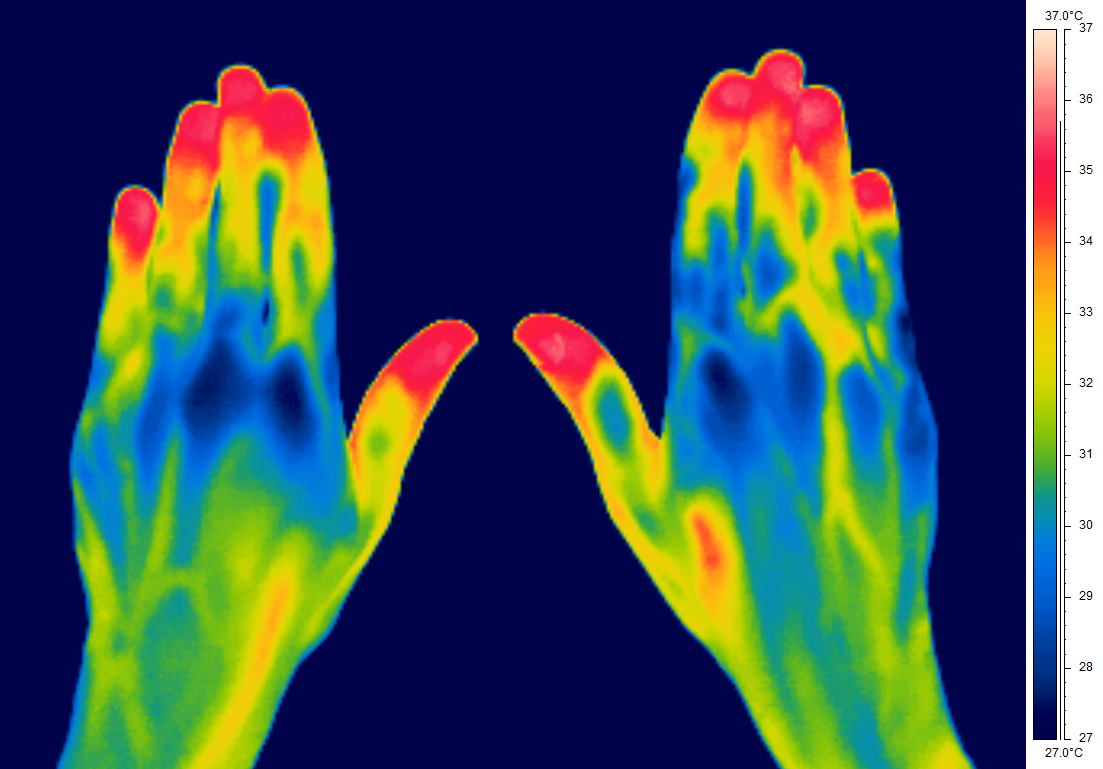
Vascular Disorders. Thermal imaging is an effective technique for detecting small temperature changes due to vascular disorders, it is non-invasive, portable, compact and safe. An article published in The British Journal of Diabetes & Vascular Disease concluded that: 'Thermography coupled with clinical findings is used for medical diagnosis of vascular disorders. Temperature gradients are observed in the affected regions of patients with vascular disorders and ischaemic gangrene, indicating abnormal blood flow in the affected region, which is well correlated with the clinical findings. The temperature in the affected regions was about 0.7-1 degrees C above the normal regions, due to slow blood circulation. The abnormalities are not observed in the same regions of healthy volunteers and normal contra-lateral limbs of the patients, demonstrating the usefulness of thermal imaging for medical diagnostics with high reliability".
Inflammation such as that caused by arthritis, soft tissue rheumatism, muscle trauma etc, produce localised areas which radiate a different amount of heat than the surrounding areas. This means that the degree of inflammation can be seen in a thermogram making it easier to visualise and monitor the effectiveness of various treatments.
Burns Assessment . Currently Burns units usually use an expensive Laser Doppler Imager to ascertain the degree of a burn sustained, this gives an indication of the healing potential and therefore the level of surgical intervention required. Tests have shown that using thermal imaging can produce results that are more accurate and easier to interpret. Further benefits are that being portable, the device can be taken to the patient, it is quick and easy to use, resulting in a much better experience for the patient.
Plastics, Rheumatology, Burns, Vascular, Haematology, Sports Injuries & Liver departments are all recognising the potential of utilising thermography and are asking for research trials to be carried out. These are ideal candidates for Medical Photography to offer their services to, extending their importance to the trust by improving patient care. Anova Technology are specialist suppliers of thermography systems to Medical Photography departments, offering packages complete with training and support.